
Microfilament Definition
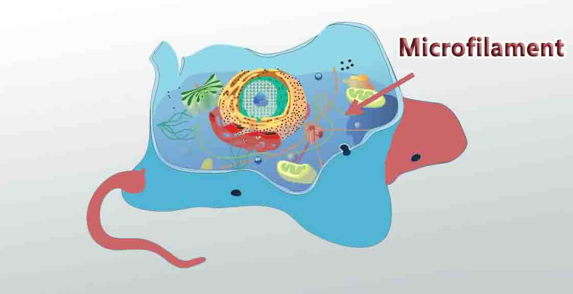
Microfilaments, also called actin filaments, are polymers of the protein actin that are part of a cell’s cytoskeleton. The cytoskeleton is the network of protein filaments that extends throughout the cell, giving the cell structure and keeping organelles in place.
Microfilaments are the smallest filaments of the cytoskeleton. They have roles in cell movement, muscle contraction, and cell division.
Microfilament Structure
Microfilaments are composed of two strands of subunits of the protein actin (hence the name actin filaments) wound in a spiral. Specifically, the actin subunits that come together to form a microfilament are called globular actin (G-actin), and once they are joined together they are called filamentous actin (F-actin).
Like microtubules, microfilaments are polar. Their positively charged, or plus end, is barbed and their negatively charged minus end is pointed. Polarization occurs due to the molecular binding pattern of the molecules that make up the microfilament. Also like microtubules, the plus end grows faster than the minus end.
Microfilaments are the thinnest filaments of the cytoskeleton, with a diameter of about 6 to 7 nanometers. A microfilament begins to form when three G-actin proteins come together by themselves to form a trimer.
Then, more actin binds to the barbed end. The process of self-assembly is aided by autoclampin proteins, which act as motors to help assemble the long strands that make up microfilaments. Two long strands of actin arrange in a spiral in order to form a microfilament.
Functions of Microfilaments
Muscle Contraction
One of the most important roles of microfilaments is to contract muscles. There is a high concentration of microfilaments in muscle cells, where they form myofibrils, the basic unit of the muscle cell. Actin is an indispensable protein for muscle movement, and microfilaments are often called actin filaments because actin is so prominent in the muscular system of the body.
In muscle cells, actin works together with the protein myosin to allow the muscles to contract and relax. Here, neither actin nor myosin can work properly without the other, and they form a complex called actomyosin. Groups of actomyosin are found in sarcomeres, the basic unit of muscle tissue.
Cell Movement
Microfilaments play a role in causing cells to move. This occurs throughout the body and it is also very important for organisms whose entire body consists of one cell, such as amoebae; without microfilaments, they would not be motile.
Actomyosin plays a role here just as it does in muscle cells. In order for cells to move, one end of a microfilament must elongate while the other end must shorten, and myosin acts as a motor to make this happen.
Microfilaments also have a role in cytoplasmic streaming. Cytoplasmic streaming is the flow of cytoplasm (the contents of the cell, including the fluid part called cytosol and cell organelles) throughout the cell.
It allows nutrients, waste products, and cell organelles to travel from one part of the cell to another. Microfilaments can attach to a cell organelle and then contract, pulling the organelle to a different area of the cell.
Cell Division
Another important function of microfilaments is to help divide the cell during mitosis (cell division). Microfilaments aid the process of cytokinesis, which is when the cell “pinches off” and physically separates into two daughter cells. During cytokinesis, a ring of actin forms around the cell that is separating, and then myosin proteins pull on the actin and cause it to contract.
The ring gets narrower and narrower around the cell, dragging the cell membrane with it, until it splits into two cells. Afterward, the microfilaments depolymerize, or break down, into actin molecules, causing the ring to dissemble when it is no longer needed.
Other Cytoskeletal Components
The two other types of filaments that make up the cytoskeleton are intermediate filaments and microtubules. Intermediate filaments are larger than microfilaments, with a diameter of about 10 nm, and microtubules are bigger than intermediate filaments at 23 nm.
Intermediate filaments bear tension in the cell, give the cell structure, and organize cell organelles and tether them in place. Microtubules have roles in transporting organelles within the cell, forming the mitotic spindle during cell division, and forming structures like cilia and flagella that help certain cells move.
Microfilaments, intermediate filaments, and microtubules all work together as part of the cytoskeleton to organize the cell and help it carry out its functions.
Related Biology Terms
- Actin – The protein that spontaneously comes together to form microfilaments.
- Cytoskeleton – A network of protein filaments that extends throughout the cytoplasm of the cell.
- Actomyosin – A complex of the proteins actin and myosin that is responsible for muscle movement.
- Cytoplasmic streaming – The flow of cytoplasm throughout the cell; it transports molecules and organelles within the cell from one place to another.